Tissues (dosma.tissues)
This page details the different tissues that are supported by DOSMA and briefly explains the methods used for analysis and visualization.
Tissues are loosely defined as structures of interest in anatomical regions. For example, both the femur and femoral cartilage are considered tissues are tissues of the knee.
DOSMA currently supports 4 tissues:
Femoral cartilage (fc)
Tibial cartilage (tc)
Patellar cartilage (pc)
Meniscus (men)
Different tissues have different quantitative profiles, visualization methods, and analysis techniques. The modules for the supported tissues implement and use these differences for analysis of each tissue.
Handles analysis and visualization for femoral cartilage. |
|
Handles analysis and visualization for tibial cartilage. |
|
Handles analysis and visualization for patellar cartilage. |
|
Handles analysis and visualization for meniscus. |
Femoral Cartilage
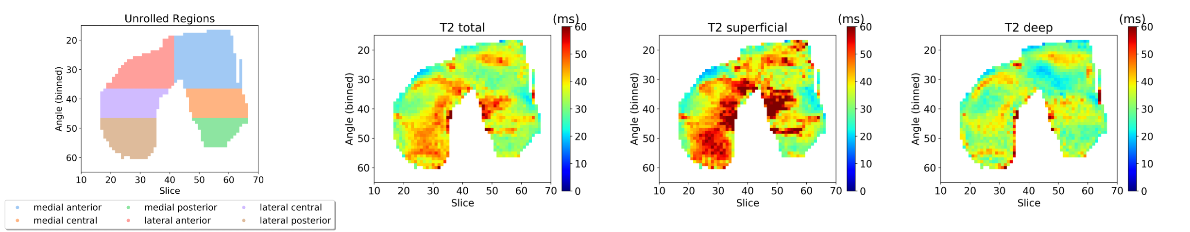
Femoral cartilage has been closely studied for evaluating knee health. The structure is often divided into sub-regions to evaluate the sensitivity of different regions of the knee to chronic diseases such as osteoarthritis. These regions are identified by three planes (12 regions):
Sagittal: Medial, Lateral
Coronal: Anterior, Central, Posterior
Depth: Deep, Superficial
For example, the deep-anterior-medial femoral cartilage tissue is one region. To analyze differences in these regions, T2 maps can be unrolled onto a 2D plane [MJS+17].
DOSMA supports automatic division of femoral cartilage into these regions and unrolling of these regions. Unrolled maps are produced for deep, superficial, and total (combined deep and superficial) layers as seen below.
Tibial Cartilage
Tibial cartilage is a flatter surface and is often divided across the three common planes:
Sagittal: Medial, Lateral
Coronal: Anterior, Central, Posterior
Axial: Deep, Superficial
DOSMA automatically divides the tissue into these regions and produces corresponding visualizations.
Patellar Cartilage
Patellar cartilage is a thin, flat tissue. Because of this structure, it is not often divided. However, there has been work that may suggest that deep/superficial differences in the patellar cartilage may be insightful.
DOSMA divides patellar cartilage into deep/superficial layers across the coronal plane.
Meniscus
DOSMA supports quantitative processing and segmentation of the mensiscus. However, visualization for the meniscus is not yet supported.